Daily from 9 a.m. to 6 p.m.
Moscow time
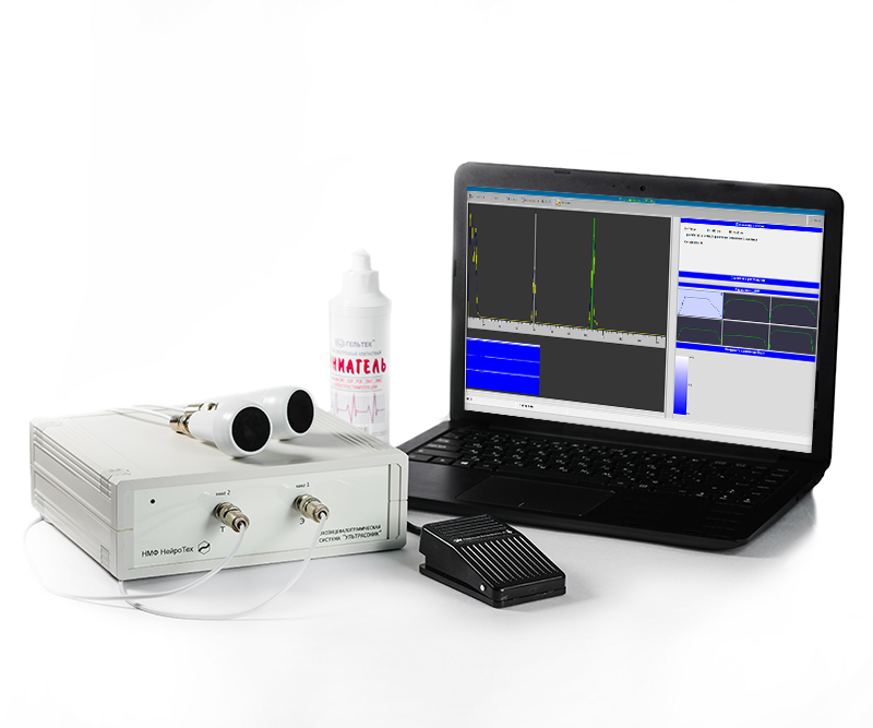
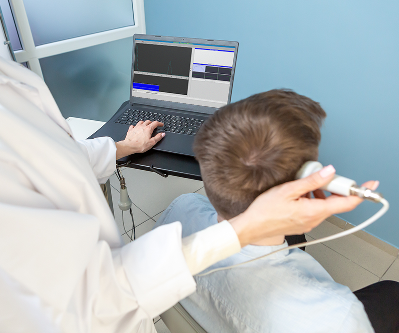
Ultrasonic Echoencephalograph (echoencephaloscope) is a classic ultrasound device designed to diagnose diseases and injuries of the brain. The difference between the portable and stationary versions is in the configuration of the system delivery, which includes a fully functional mini-laptop and a special case for easy movement with the system.
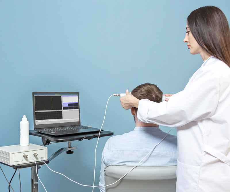
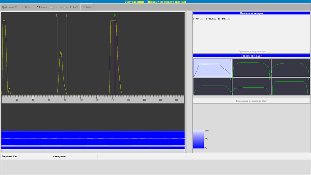
Computerized Ultrasonic echoencephaloscope for diagnosing brain disorders and injuries is designed to conduct a one-dimensional echoencephalographic examination of the brain using generally accepted methods.
The system allows for diagnostics of conditions of the brain and intracranial vessels in order to identify brain disorders and injuries using echo-pulse location methods. The complex can be effectively used in emergency and planned neurology, traumatology, resuscitation, neurosurgery, etc.
The device is connected via a USB interface to a mini-laptop, onto which the software is installed that implements generally accepted methods for measuring and evaluating the main parameters and identifying pathological changes in the brain.
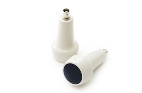
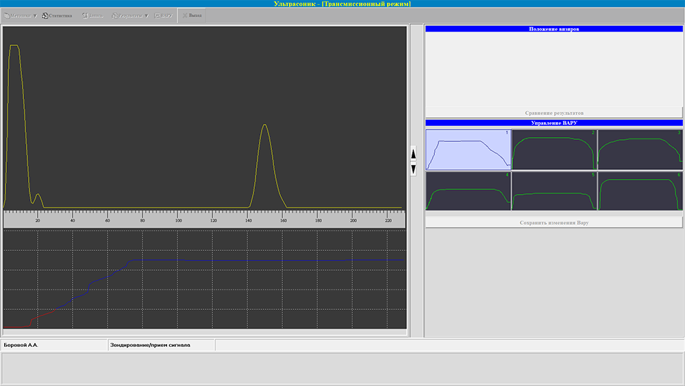
The device uses two sensors: one for emission modes, the other for transmission mode.
Computerized Ultrasonic echoencephaloscope for diagnosing brain disorders and injuries allows to:
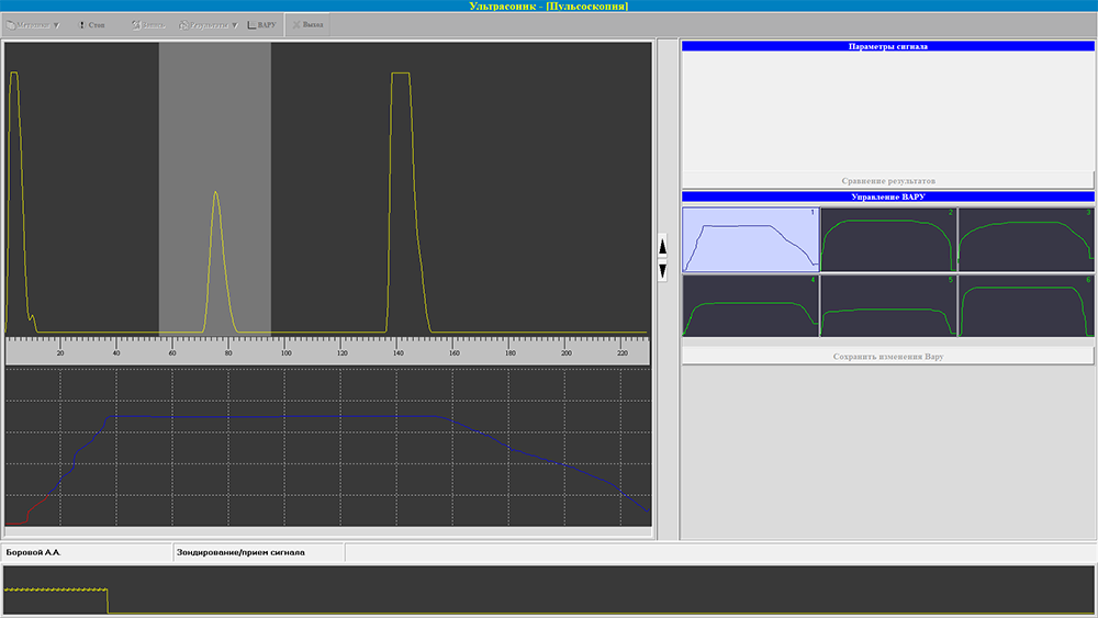
The echoencephaloscope is equipped with a software-controlled automatic temporal gain control (TGC), which enables the recording of high-quality echo signals.
A probing depth of 230 mm with a “dead” zone of no more than 15 mm allows for the identification of pathological formations in the periosteal cranial space.
Ultrasonic echoencephaloscope has the following unique features:
Software functions:
|
Probing depth |
230 mm min |
|
Dead zone length |
40 mm max |
|
Longitudinal resolution |
1 mm max |
|
Linear dimension measurement range (on screen) |
1 – 230 mm |
|
Limits of permissible error of the device when measuring linear dimensions |
±(0.03L*+1) mm max |
|
Power supply |
(220±22) V, frequency (50±0.5) Hz |
|
Power consumed by the device from the AC network |
10 W max |
|
Overall dimensions of the device housing |
225 x 220 x 72 mm |
|
Device weight |
2 kg max |
|
Time to establish the operating mode of the device (excluding the time it takes to turn on the computer) |
10 sec max |
|
Average service life of the device before decommissioning |
at least 5 years with an average intensity of use of 5 hours per day |
| Аппаратный блок «Ультрасоник» | 1 pc. |
| Cable for connection to PC (USB cable, type A-B) | 1 pc. |
| Device power cable (Europlug) | 1 pc. |
| Ultrasonic sensor | 2 pcs. |
| Cable for connecting ultrasonic sensors | 2 pcs. |
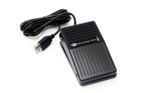
| Pedal | 1 pc. |
| Mobile PC (like a netbook) | 1 pc. |
| Computer mouse | 1 pc. |
| Case for system carrying | 1 pc. |
| Ultrasonic gel | 1 pc. |
| Flash memory stick with software | 1 pc. |
| Operational documentation | 1 set |
This site is protected by reCAPTCHA and the Google Privacy Policy and Terms of Service apply
This site is protected by reCAPTCHA and the Google Privacy Policy and Terms of Service apply


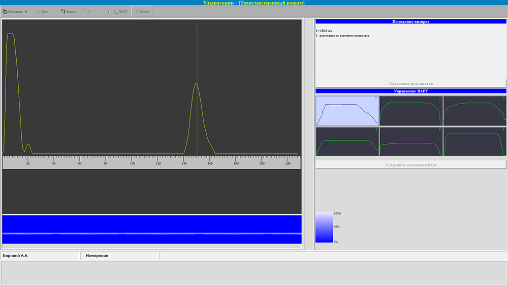
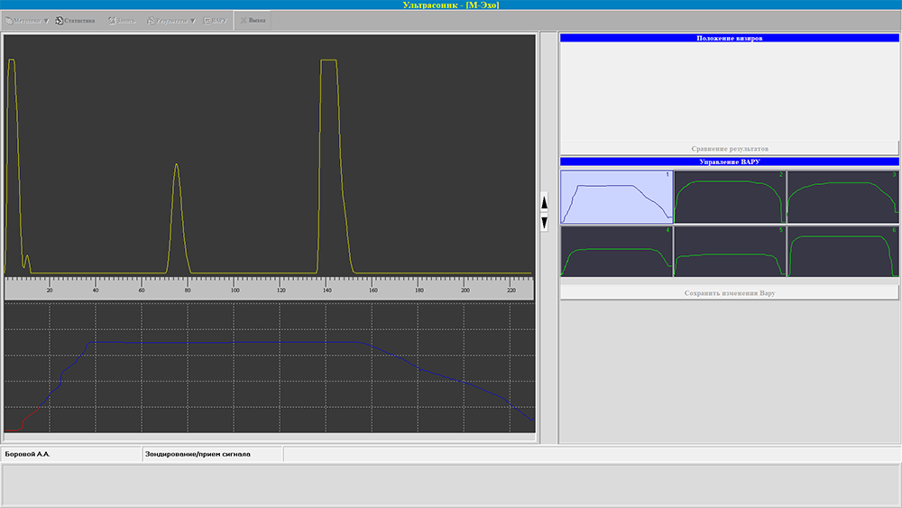
Ultrasonic is equipped with software-controlled temporal gain control (TGC), which ensures high quality of echo signals
Ultrasonic Echoencephalograph enables to quickly assess the presence of a brain tumor and hematoma
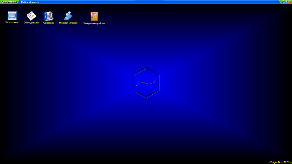
The user-friendly and intuitive database interface enables to quickly fill in patient information during registration in the electronic card file
Optimized manufacturing process and careful technical control ensure high quality and affordable price of the equipment
Введите номер телефона и мы Вам перезвоним в ближайшее время!
Ваш запрос принят!
С вами свяжутся в ближайшее время.
This site is protected by reCAPTCHA and the Google Privacy Policy and Terms of Service apply